A New Hertel et al. Publication in mBio!

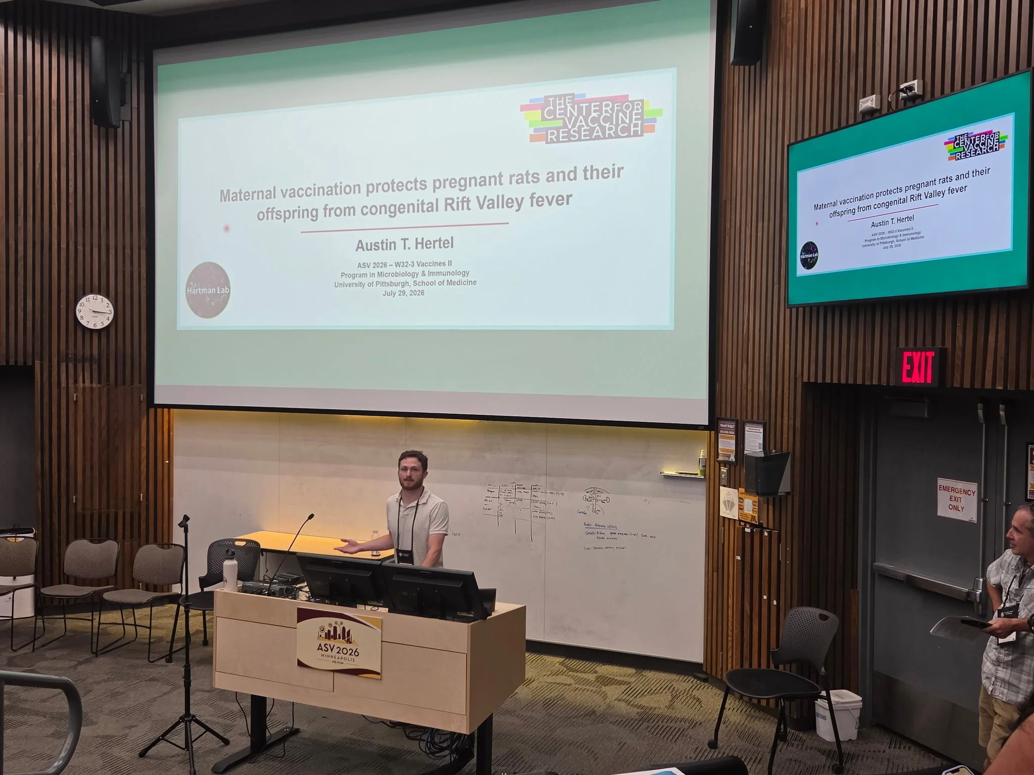
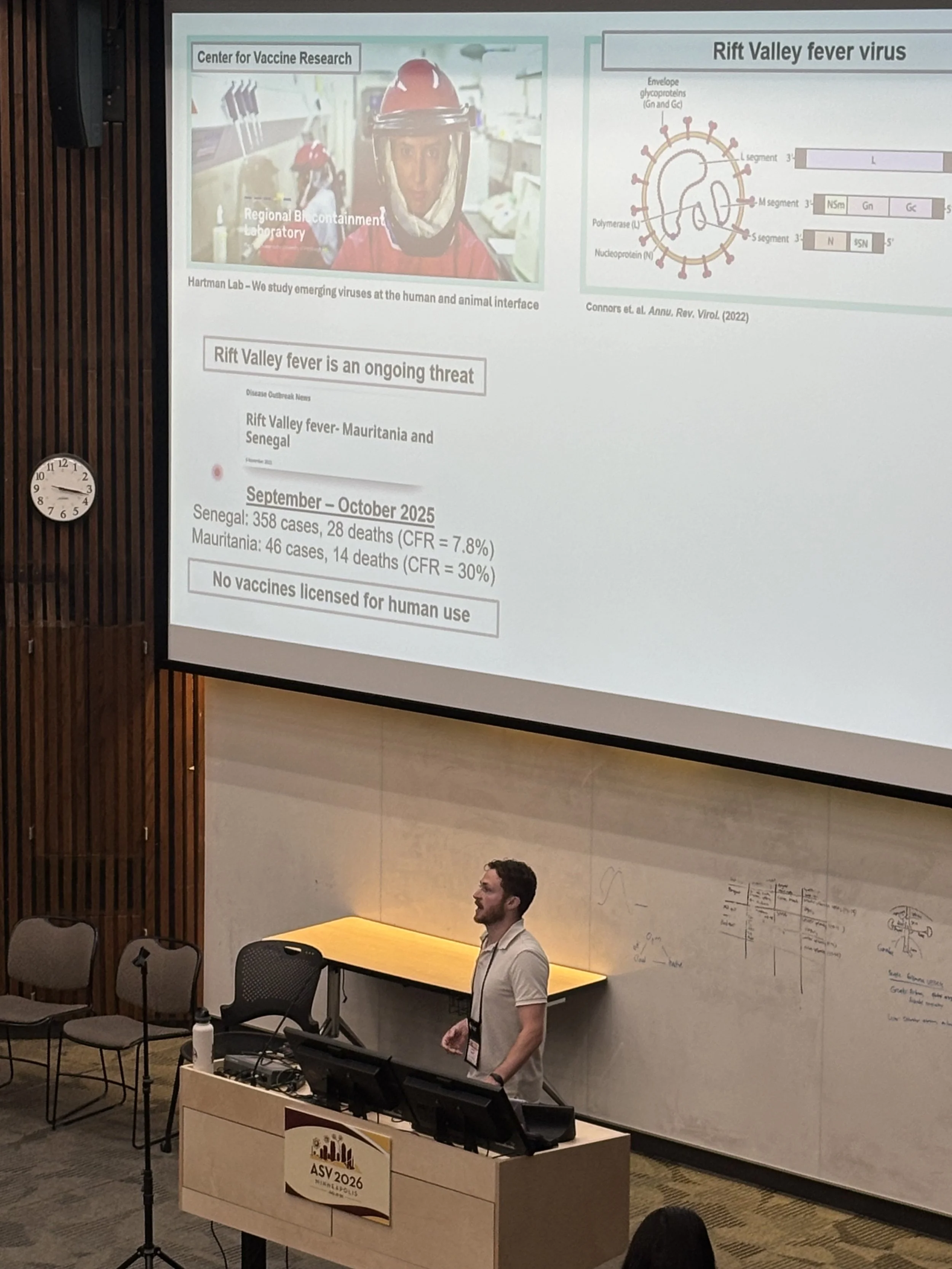
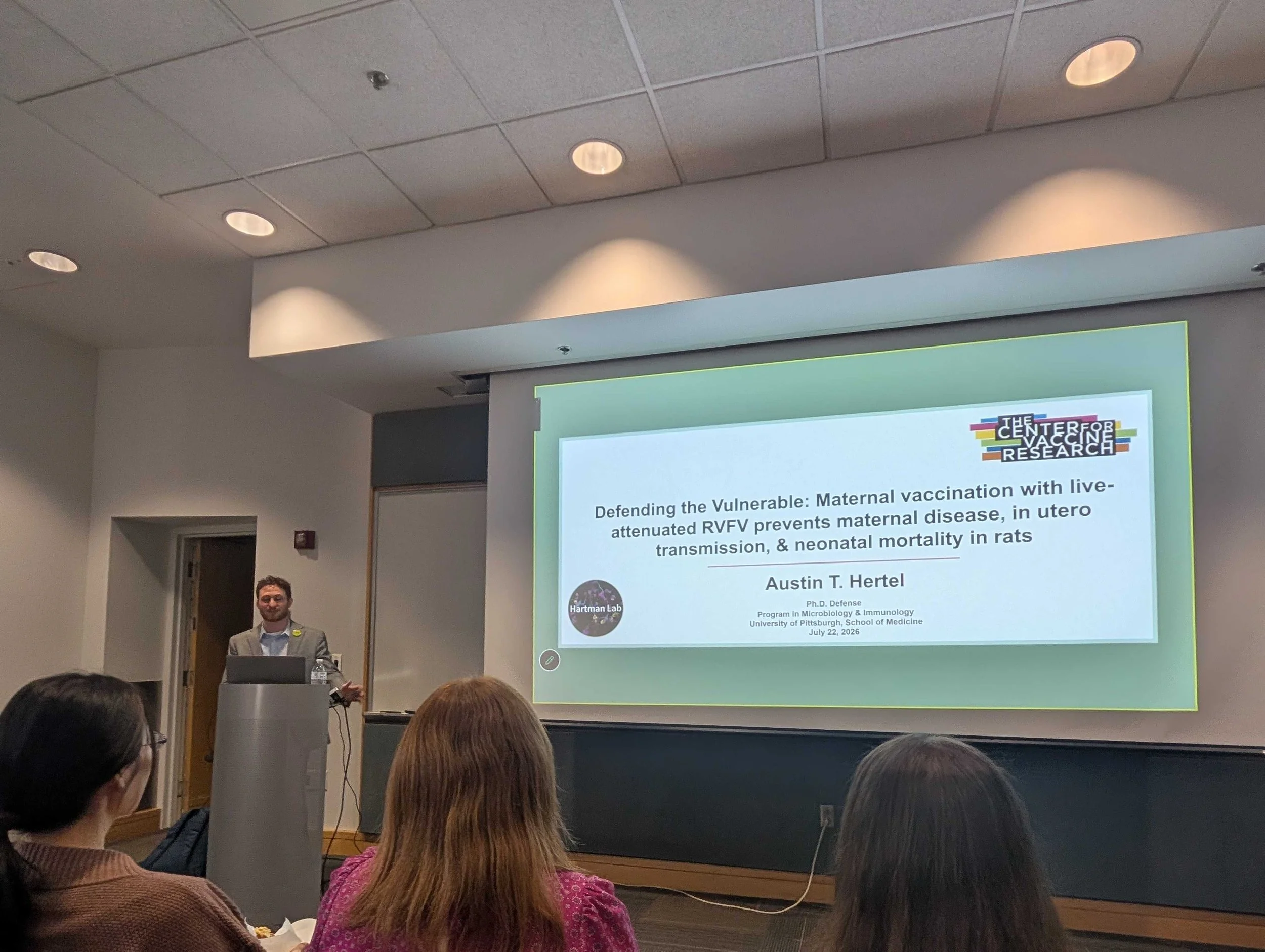
Our very own Austin Hertel’s 2nd publication as first author was just made available in the American Society for Microbiology’s mBio! “Maternal vaccination protects dams and prevents in utero transmission of Rift Valley fever virus in rats,” focuses on vertical transmission of Rift Valley fever virus (RVFV) between mother and fetus in rats, specifically evaluating the safety and protective ability of a live-attenuated RVFV vaccination in dams.

RVFV causes spontaneous fetal loss among domesticated livestock populations, and has also been shown to cause fetal loss and hepatic disease in human infants born to pregnant individuals infected with RVFV. The disease poses a serious threat to vulnerable pregnant populations, especially without licensed vaccines currently available for human use. Previously developed live-attenuated vaccines retained partial virulence causing fetal death and congenital defects and thus are not suitable for human use. The vaccine used in this study is a live-attenuated version of the ZH501 strain of RVFV which has been generated by reverse genetics to lack the virulence genes encoding the non-structural proteins NSs and NSm. Vaccination with RVFV-delNSs/Nsm protects mother and fetus from wild-type viral challenge while also being avirulent.

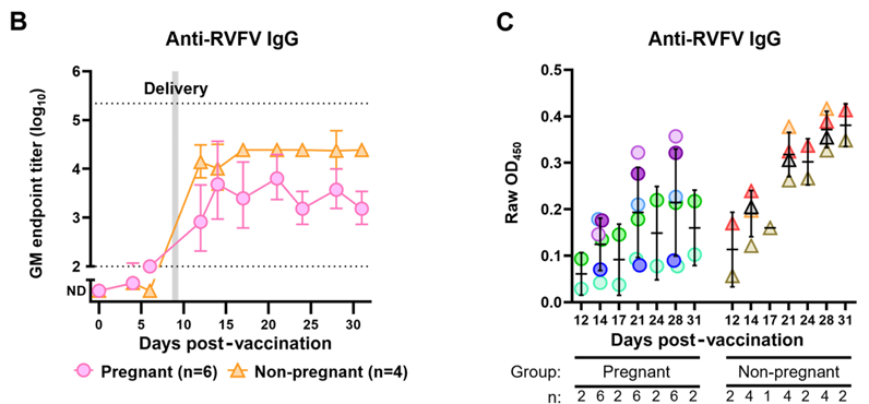
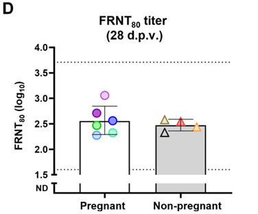
Immune response titers demonstrated pregnant and non-pregnant rats responded similarly to vaccination with RVFV-delNSs/NSm. Titers of RVFV-specific IgG1 and IgG2a in pregnants rats vaccinated with the live-attenuated RVFV strain indicated the Th2- and Th1-mediated responses of rats are similar regardless of pregnancy status. There were some slight differences in IgG response, although neutralization capacity between pregnant and non-pregnant rats were comparable.

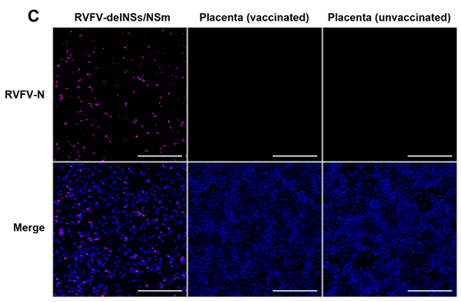
Immunofluorescent images of Vero E6 cells inoculated with placental homogenate.

The live-attenuated RVFV vaccine also spreads minimally throughout the pregnant dam when vaccinated during early gestation at a high dose (1 x 10^5 PFU of RVFV-delNSs/Nsm). Live virus could not be isolated from placental homogenates of two pregnant dams sacrificed prior to giving birth. Two other pregnant dams vaccinated during early gestation delivered normal litter sizes with no apparent congenital abnormalities. These results indicate that there is limited clinical impact on the health of offspring when pregnant individuals receive the live-attenuated vaccine during gestation even at high doses.

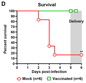
We also demonstrated vaccinated pregnant animals survive challenge from RVFV after vaccination with the live-attenuated RVFV-delNSs/NSm and sufficiently protect fetuses from in utero transmission of RVFV. Surviving animals had very low levels of vRNA detected in maternal and placental samples, with vRNA found in the fetal viscera. Infectious virus was also not found in maternal, placental, or fetal samples from these vaccinated animals indicating vaccination reduced viral burden and in utero transmission, protecting both dam and pup from RVFV clinical disease.

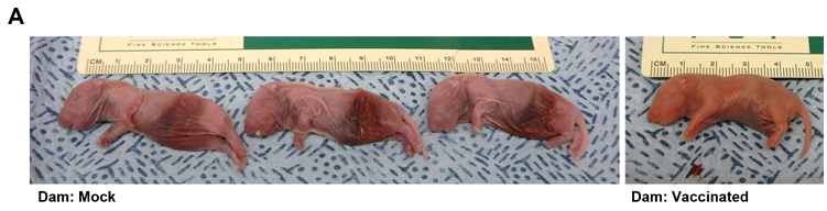
Mock-vaccinated dams delivered pups with visually apparent visceral hemorrhage and lower birth weights in comparison to vaccinated dams after surviving viral challenge. 3 of the 8 delivered pups from the lone surviving mock-vaccinated dam had this congenital abnormality while vaccinated dams that survived viral challenge delivered largely healthy litters with only 4 pups out of 70 being found dead at delivery. Viral RNA in the fetal viscera of vaccinated dams was also minimal especially in comparison to the high levels of vRNA in the fetal viscera of the mock-vaccinated dams.

Vaccination prior to pregnancy was also shown to protect dams and fetuses from RVF during future pregnancy, with female rats paired with males 10 days post vaccination that became pregnant surviving viral challenge with wild-type RVFV while all mock-vaccinated dams succumbed to lethal infection.

All in all, these findings display robust evidence for the safety and efficacy of the RVFV-delNSs/NSm in pregnant animals. This work provides a useful platform and building block for the use of the live-attenuated RVFV strain as a safe and viable vaccine platform for pregnant individuals. Incredible work, Austin!

CVR Trainee Day 2026

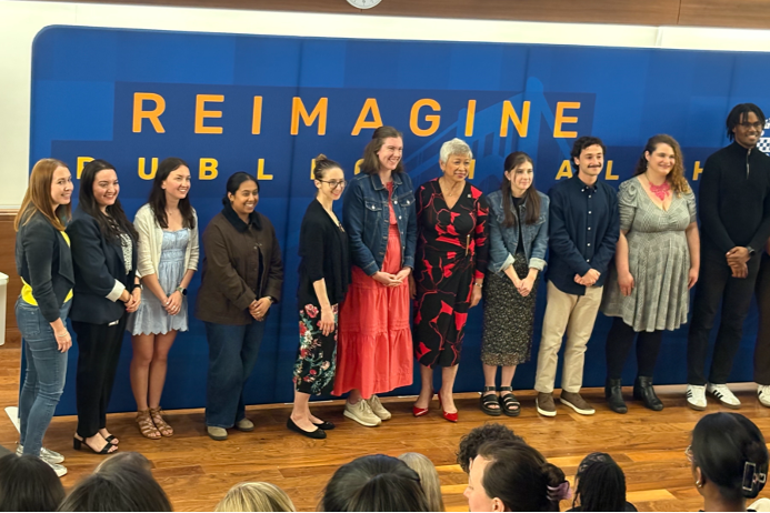
On Friday, June 5th the CVR held its fourth annual Trainee Day! The festivities of this year’s presentations were managed in-part by our very own Cade Sterling and Elon Holmes (grad students)!

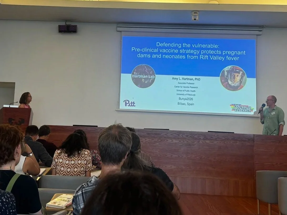
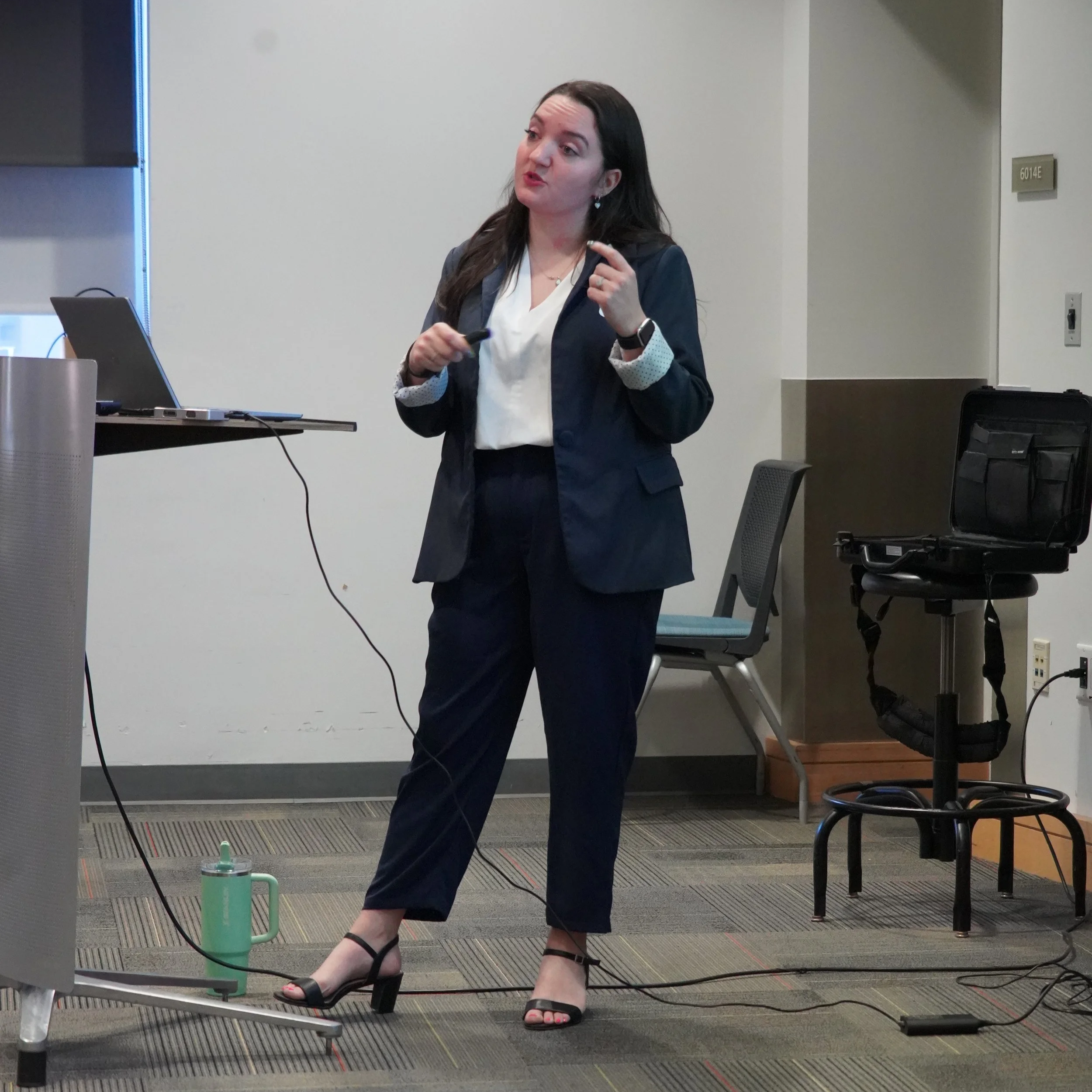
Our superstar post doc Dr. Rachael Rush opened the slate of talks for the day with a bang, presenting her work “Translational approaches to combating peribunyaviruses: the Revampp strategy.

Rachael, pictured above casually hitting an astonishing pose while presenting to a room full of distinguished peers.

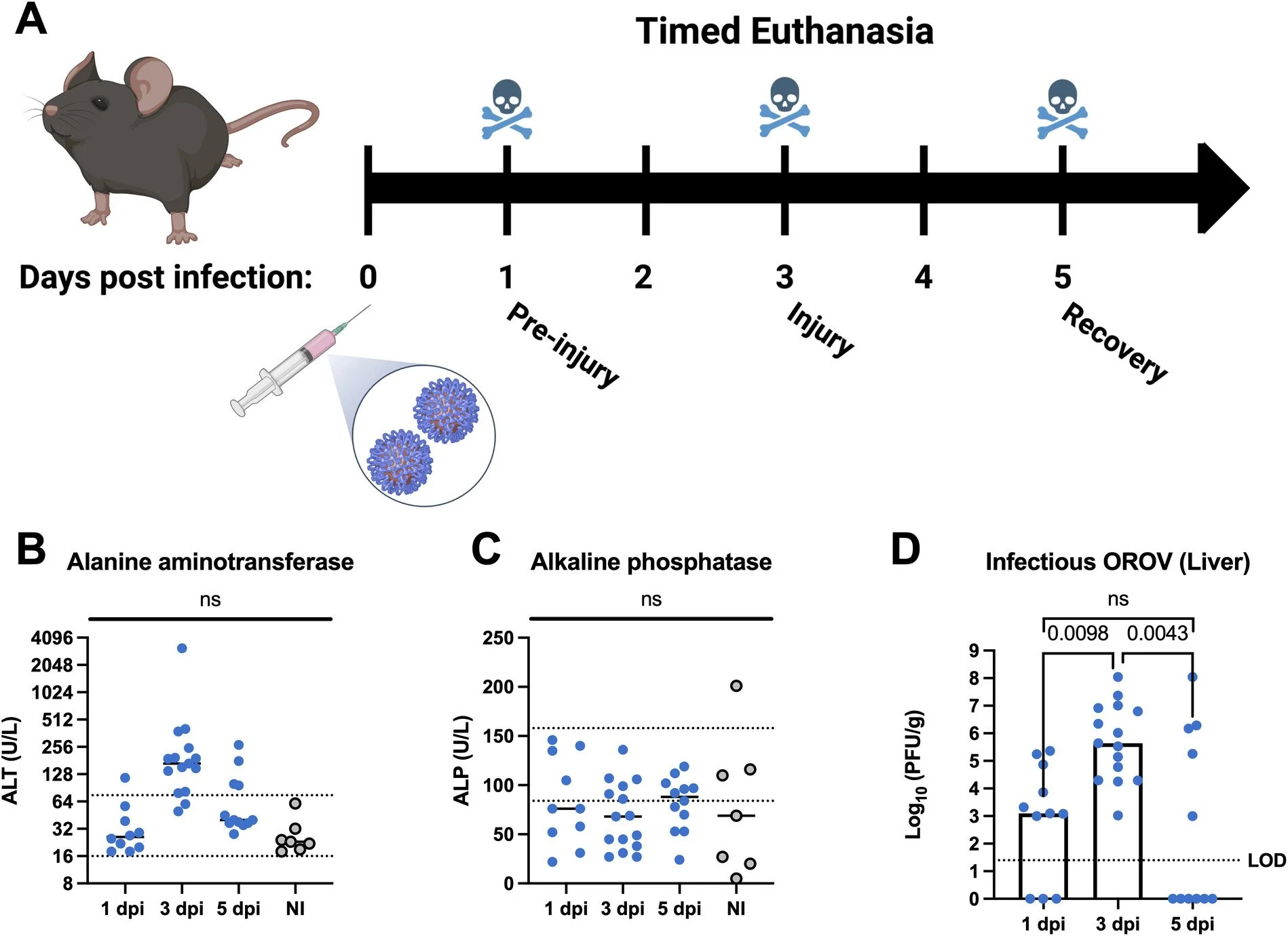
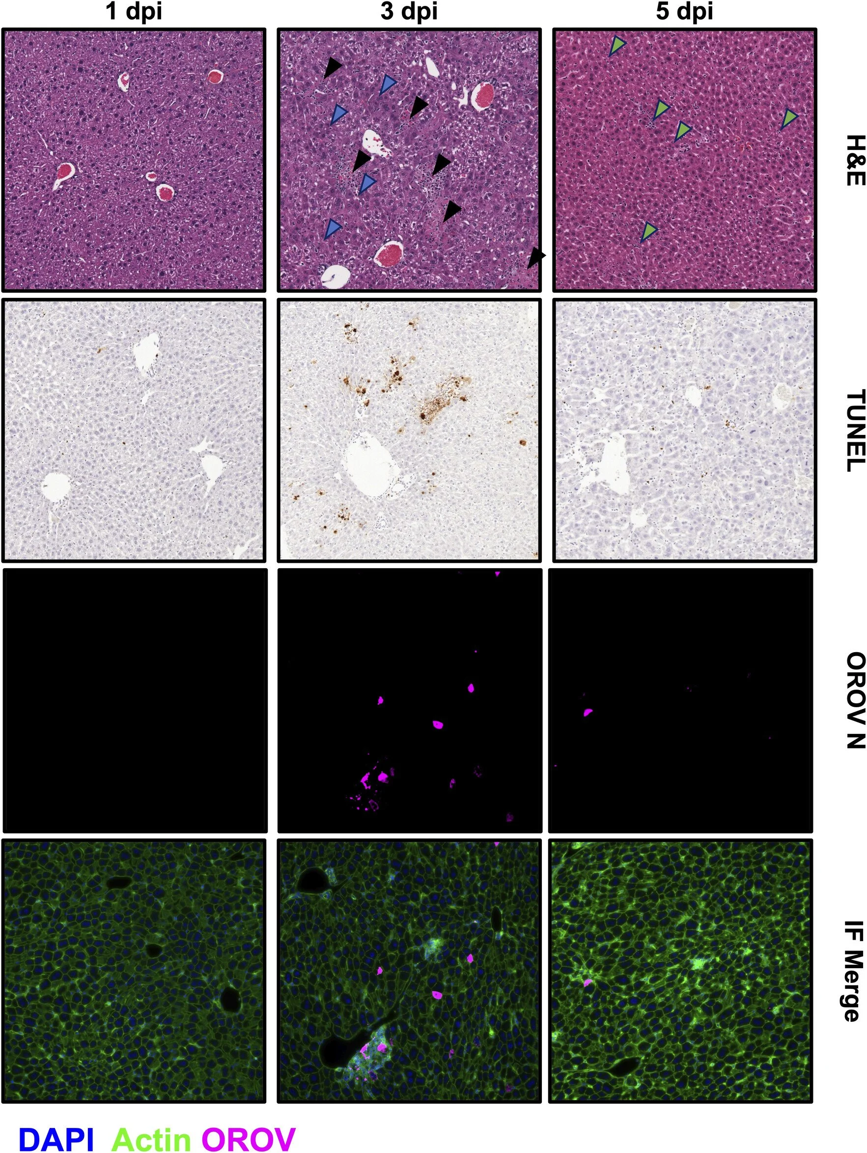
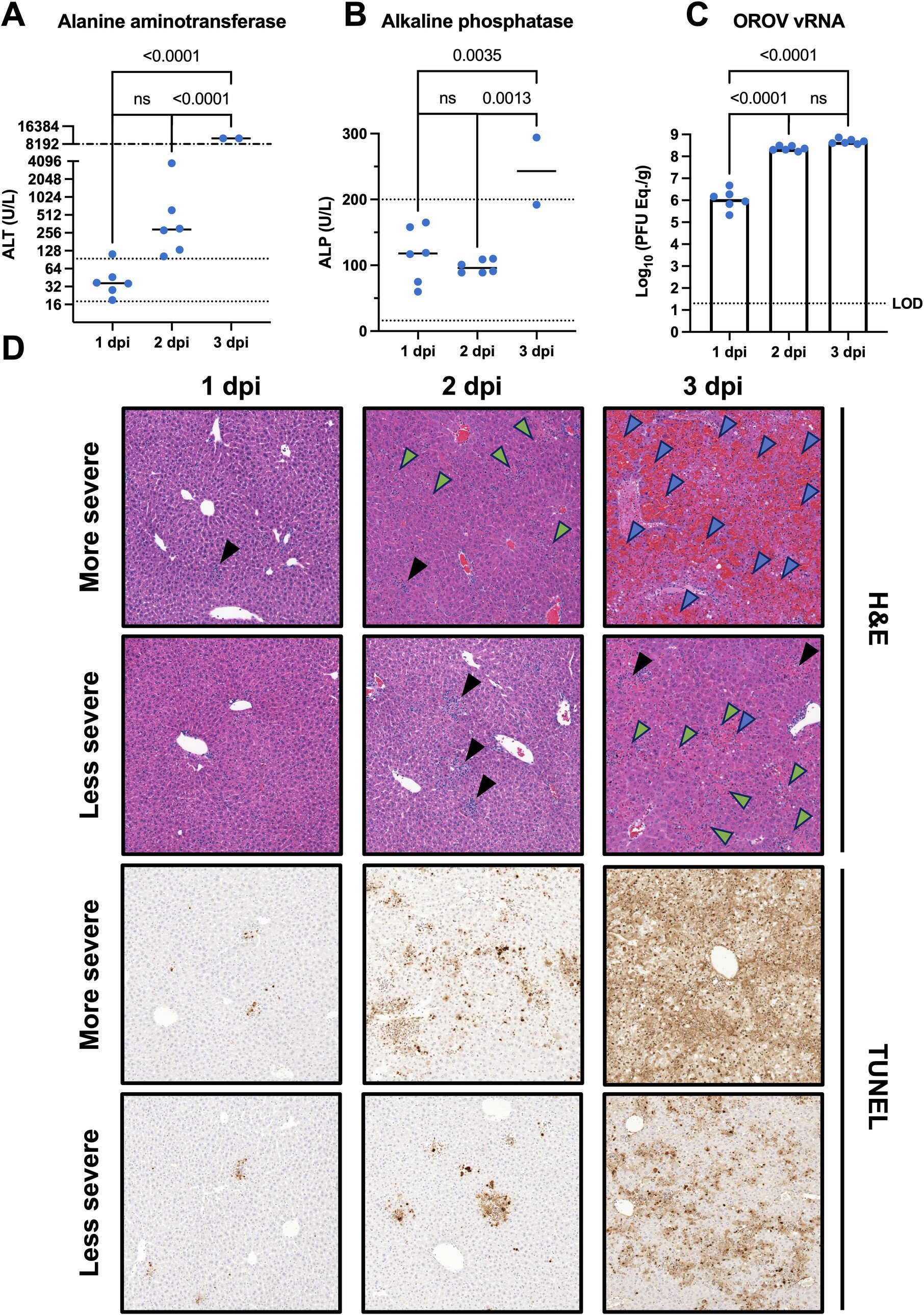
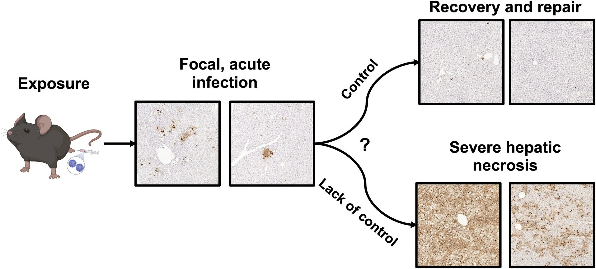
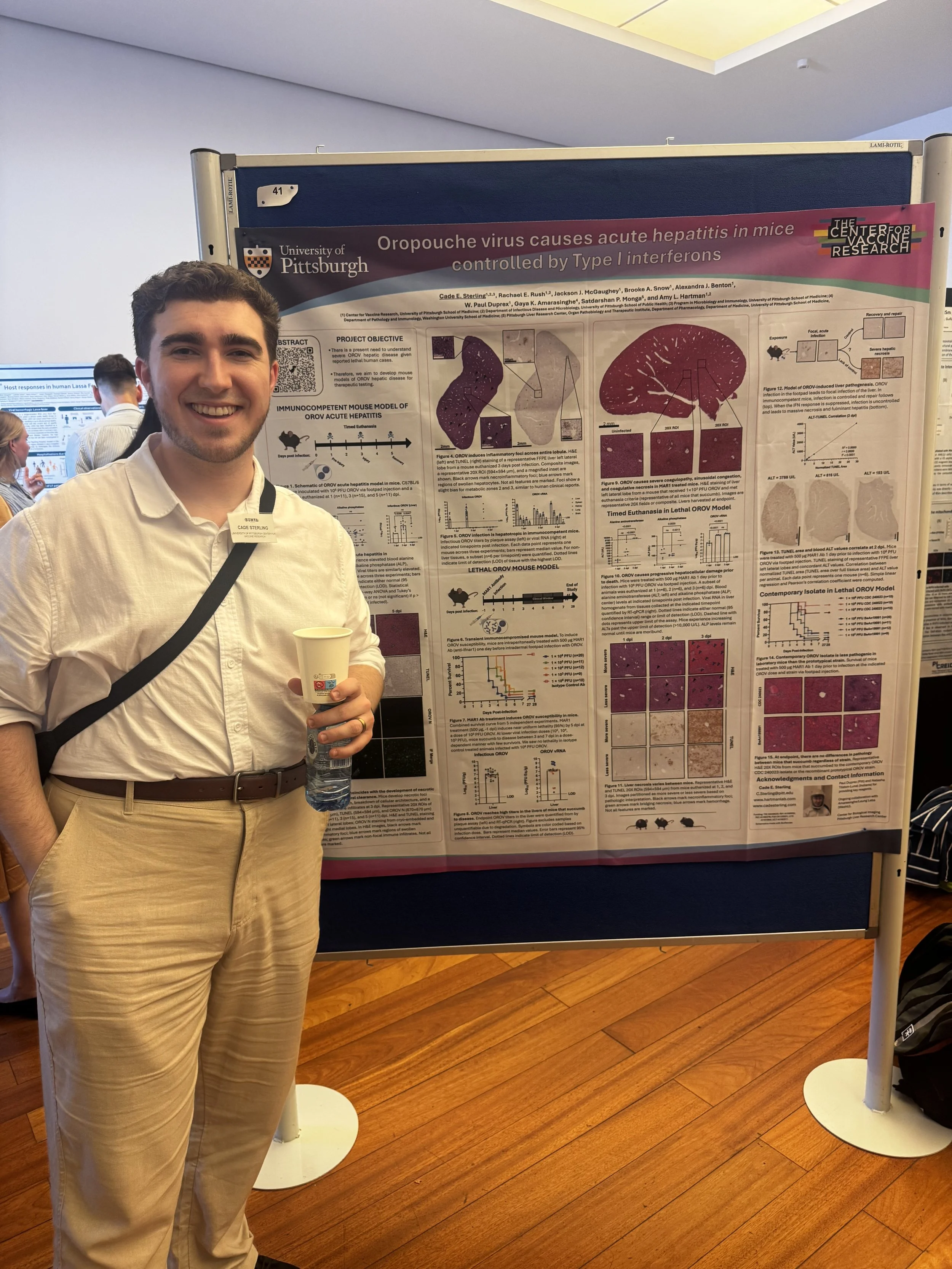
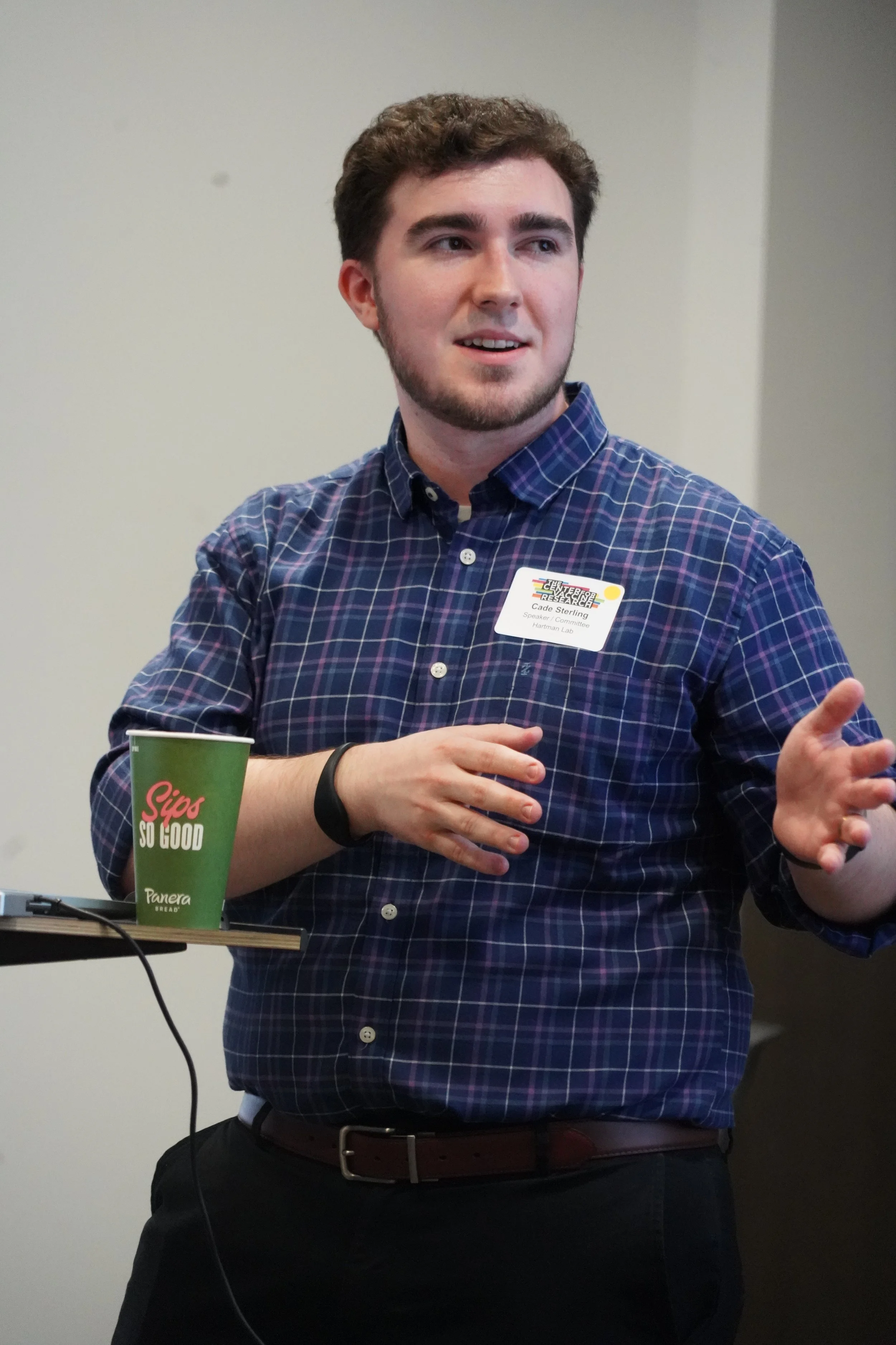
In between running around the 6th floor keeping things in line, our PhD candidate Cade also had time to deliver a fantastic presentation of his work, entitled “Oropouche virus causes acute hepatitis in mice controlled by Type I interferons.

A dapper Mr. Sterling gesturing to his slides (not shown).

Former master’s student, current Master of Science and lab tech Andrew Wheeler also presented at Trainee Day! He shared his project, “Comparing virulence of historical and contemporary Oropouche strains in the context of pregnancy.”

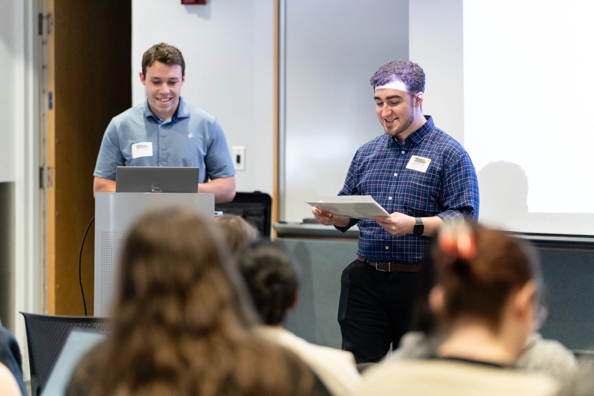
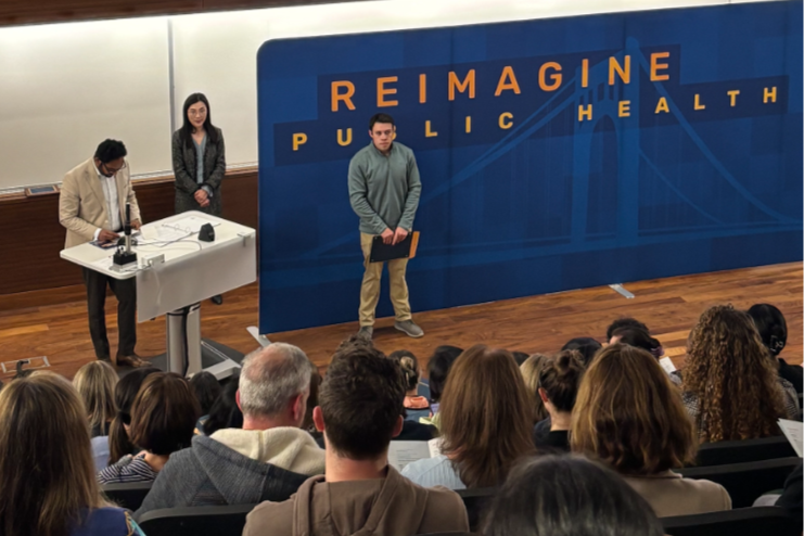
Master Wheeler, being introduced by Cade!
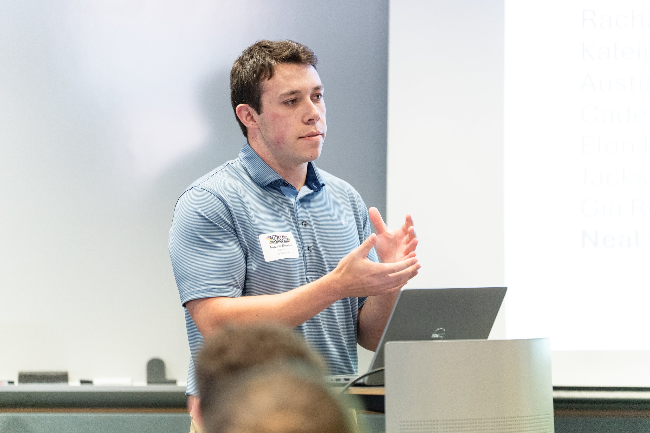
A pensive, distinguished Andrew fielding a question from the audience.

Rachael, Cade, and Andrew did a tremendous job representing the Hartman Lab and contributed to another wonderfully successful Trainee Day!

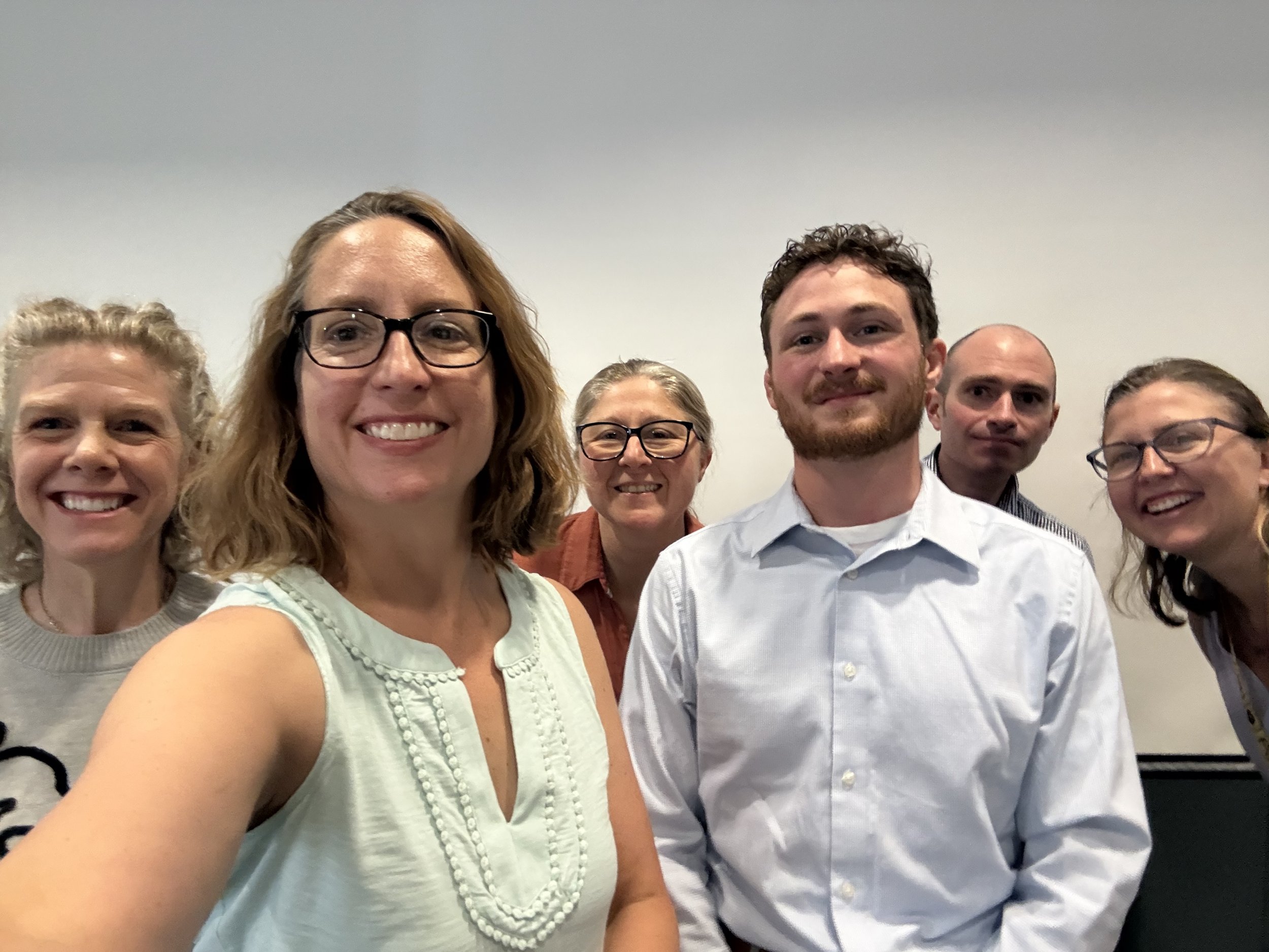
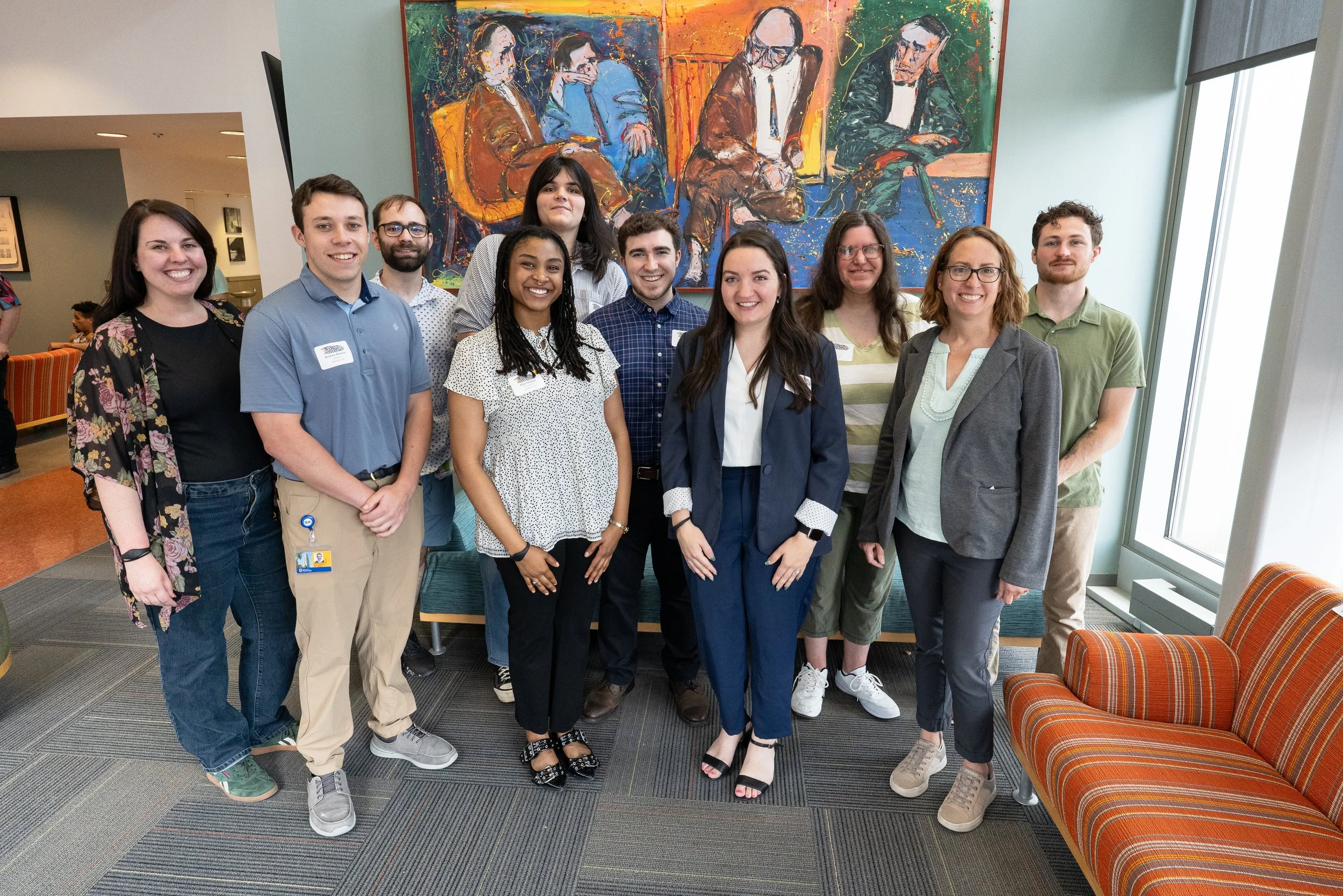
An updated lab family photo for the record books!

Graduations and New Beginnings

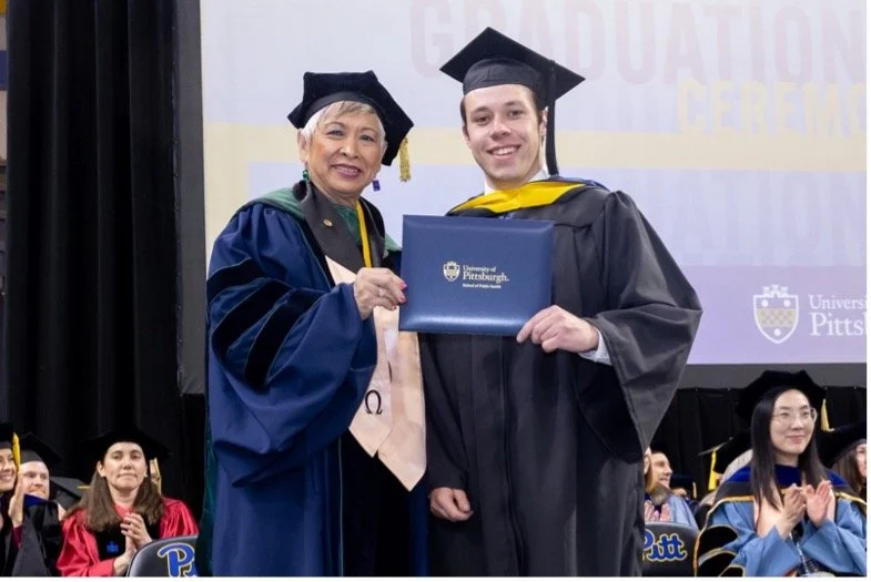
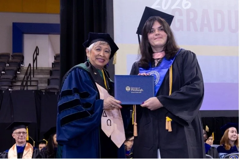
At the beginning of this month two members of our lab family successfully completed their degree programs! Andrew Wheeler graduated from Pitt’s Department of Infectious Diseases and Microbiology as a Master of Science after successfully defending his thesis on work he completed within the lab. Gia Romano (yours truly) also graduated summa cum laude from Pitt’s School of Public Health as a Bachelor of Science in Public Health with an Honors Distinction

We are also excited to announce that both Andrew and Gia will continue working with the Hartman Lab post-graduation as Research Technicians, building upon the superb work they completed as students with us. Please join us in congratulating them both!


Written by Gia Romano

A Dazzling Dean’s Day for the Hartman Lab!

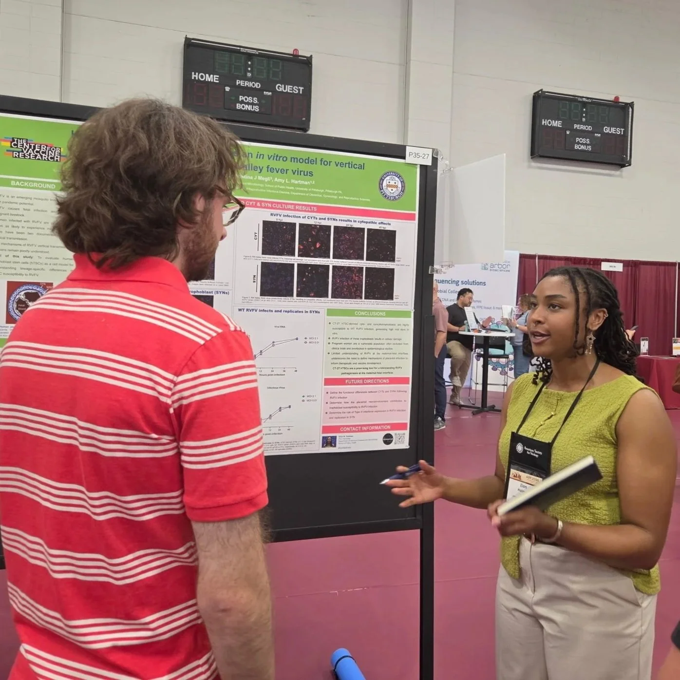
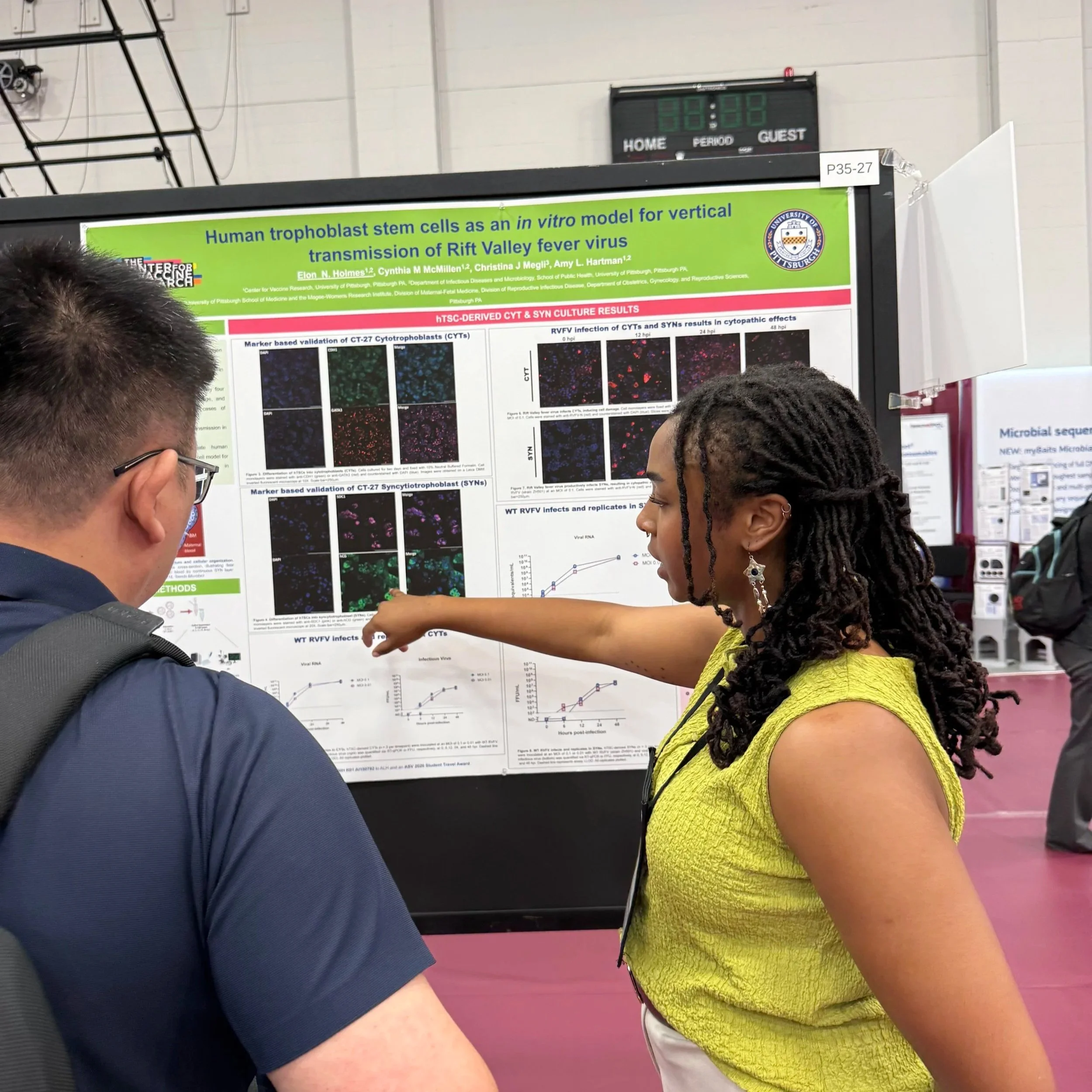
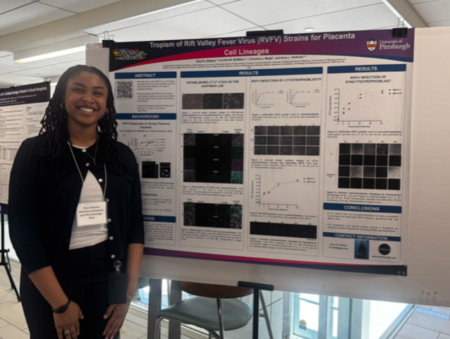
This year’s Dean’s Day at the School of Public Health was a smashing success for the Hartman Lab! The brilliant Elon Holmes presented Tropism of Rift Valley Fever Virus (RVFV) in Placenta Trophoblast Lineages highlighting her work as a PhD student collaborating with Dr. Cindy McMillen on studying the mechanisms of RVFV vertical transmission.

Elon and her fabulous poster!

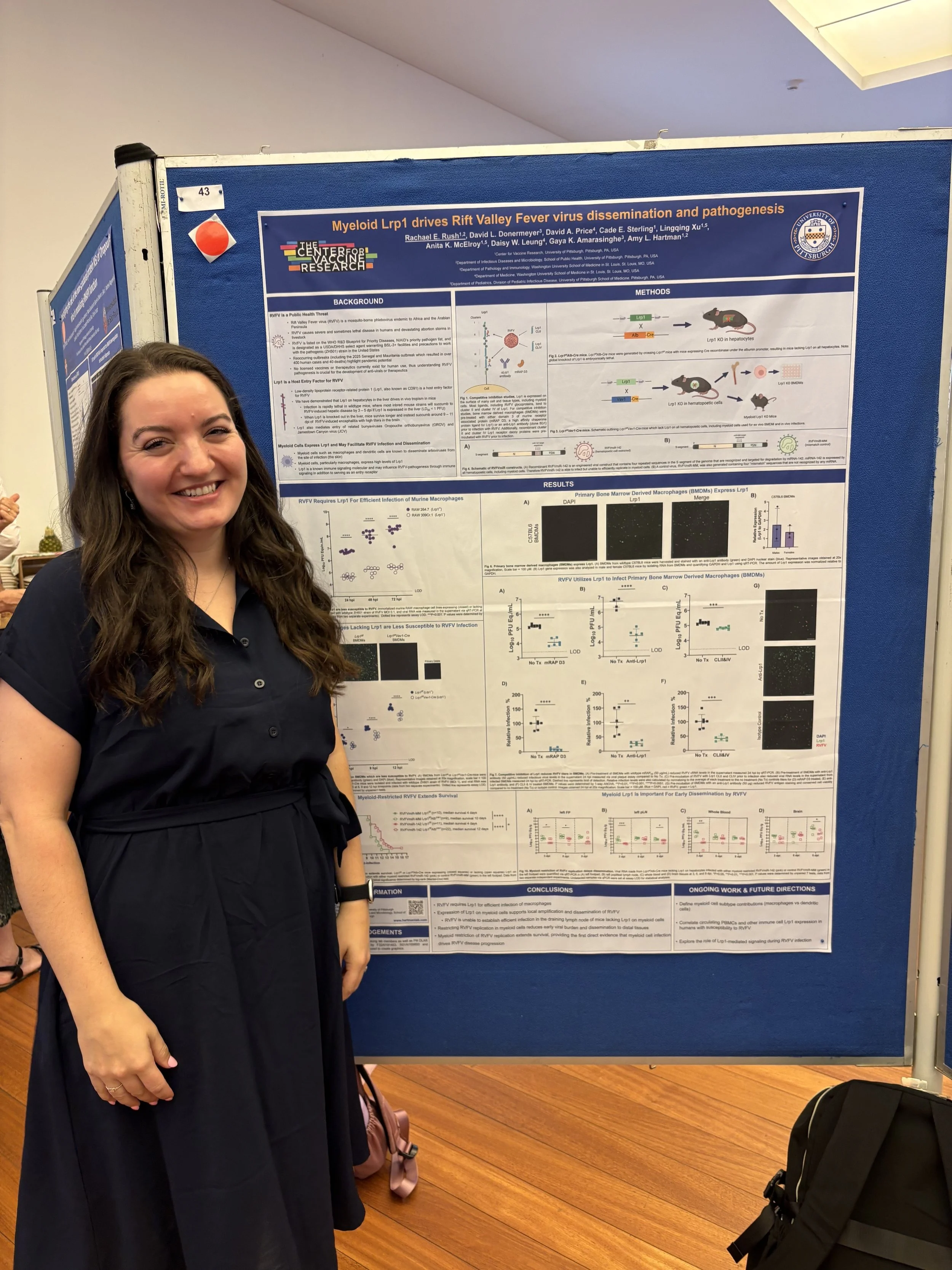
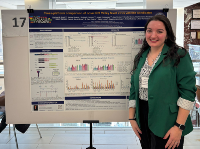
Our lovely postdoc Dr. Rachael Rush also presented Cross-Platform Comparison of Novel Rift Valley Fever Virus Vaccine Candidates discussing some of her ongoing work with the ReVAMPP project.

The wonderful Dr. Rush presenting her work!

For her exceptional work, Dr. Rush also was honored with the “Outstanding Postdoctoral Trainee Award” at Dean’s Day!

The star quality of one of our Master’s Students, Andrew Wheeler, was also highlighted as he received the “Dean’s Day Service Award”! Congratulations to Elon, Rachael, and Andrew on their incredible work and representation of the Hartman Lab!


Written by Gia Romano

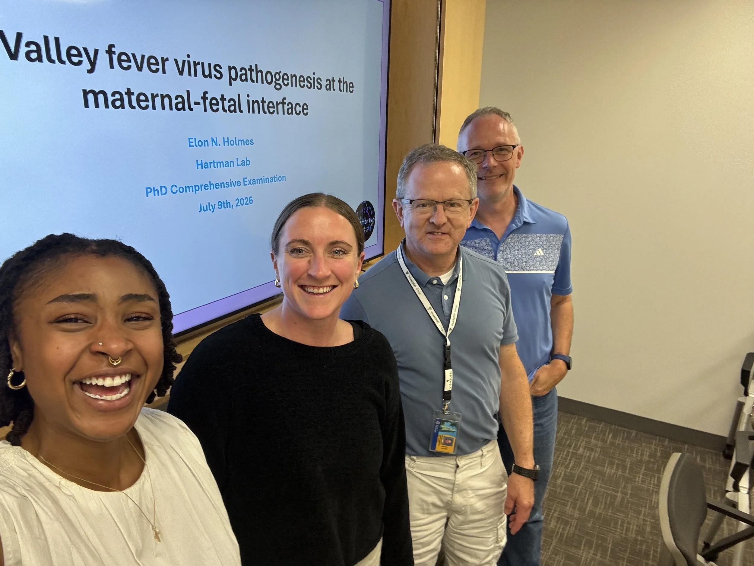
Congratulations to Master of Science, Andrew Wheeler!

We are thrilled to share that our very own Andrew Wheeler has successfully defended his thesis and become a Master of Science! Andrew’s work in our lab as a Master’s student in the Department of Infectious Diseases and Microbiology has culminated in his thesis, “Understanding host and virus factors that dictate vertical transmission of emerging bunyaviruses.”
Andrew defending his thesis, and looking spiffy in the process!
Andrew primarily worked alongside Dr. Cindy McMillen with support from our undergraduate student Neal Gupta to compare the tropism and pathologies of bunyaviruses within host placental trophoblasts.

Andrew successfully defended his thesis on Thursday, April 16th and will continue working with us at the Hartman Lab as a Research Technician this spring after graduation this May! We are so proud of the work he has already accomplished and will continue to do as a member of our lab family.

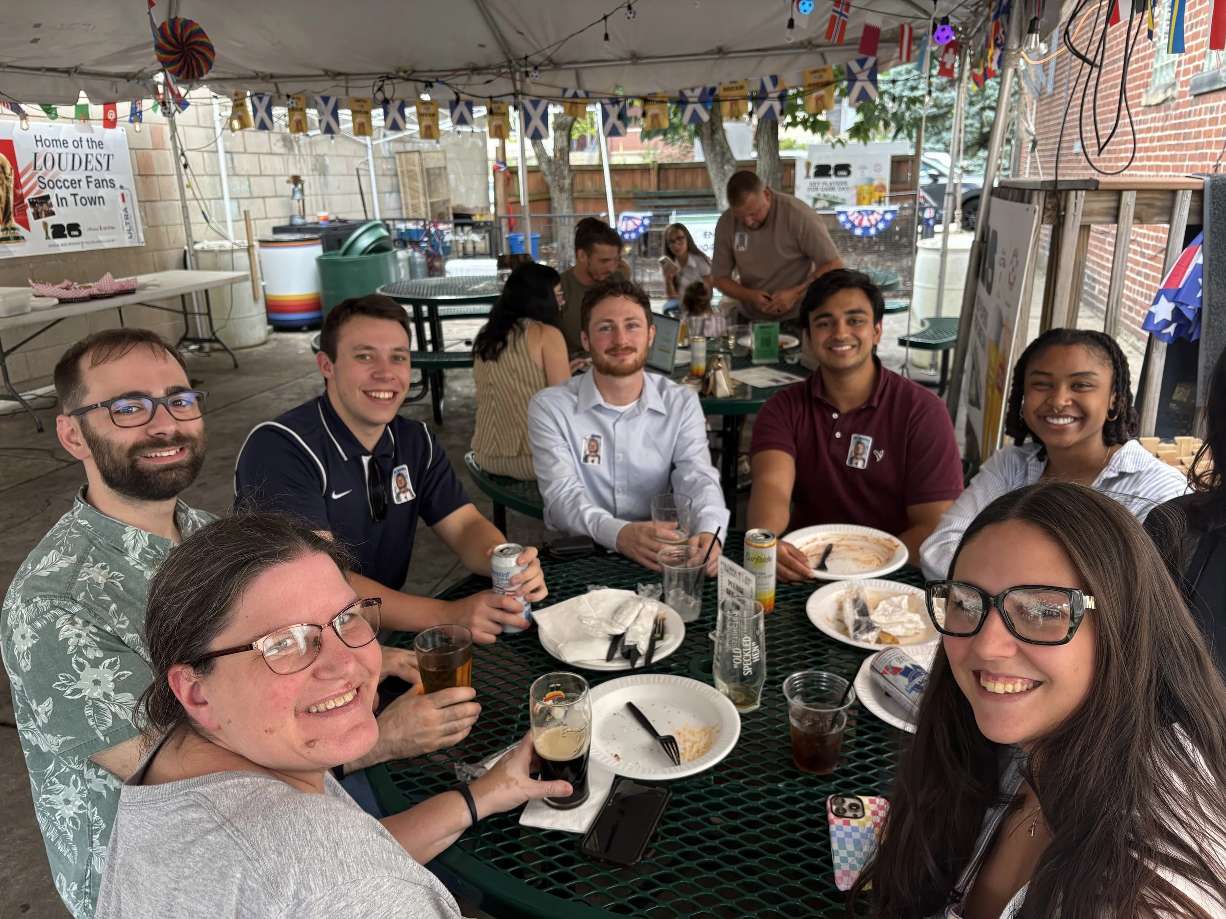
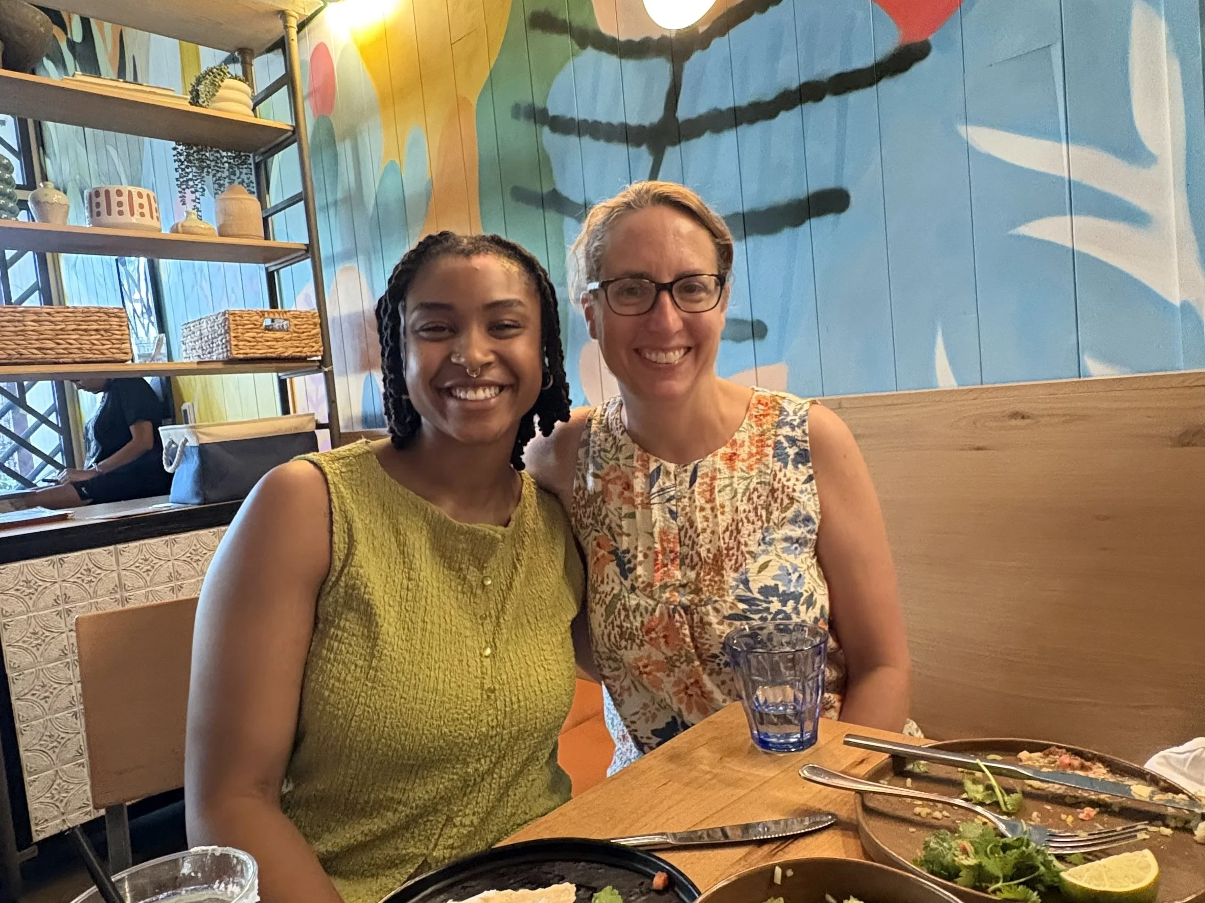
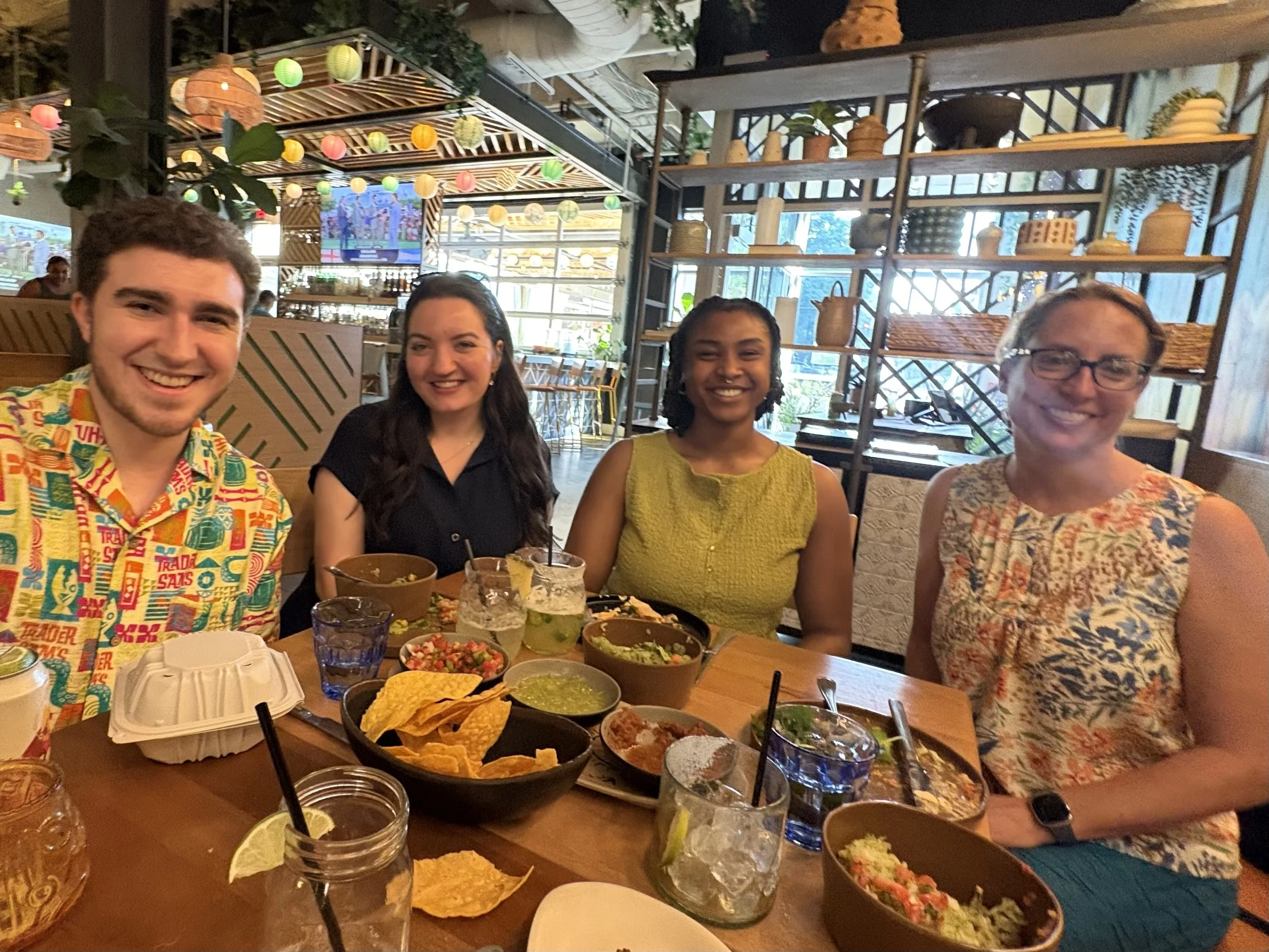
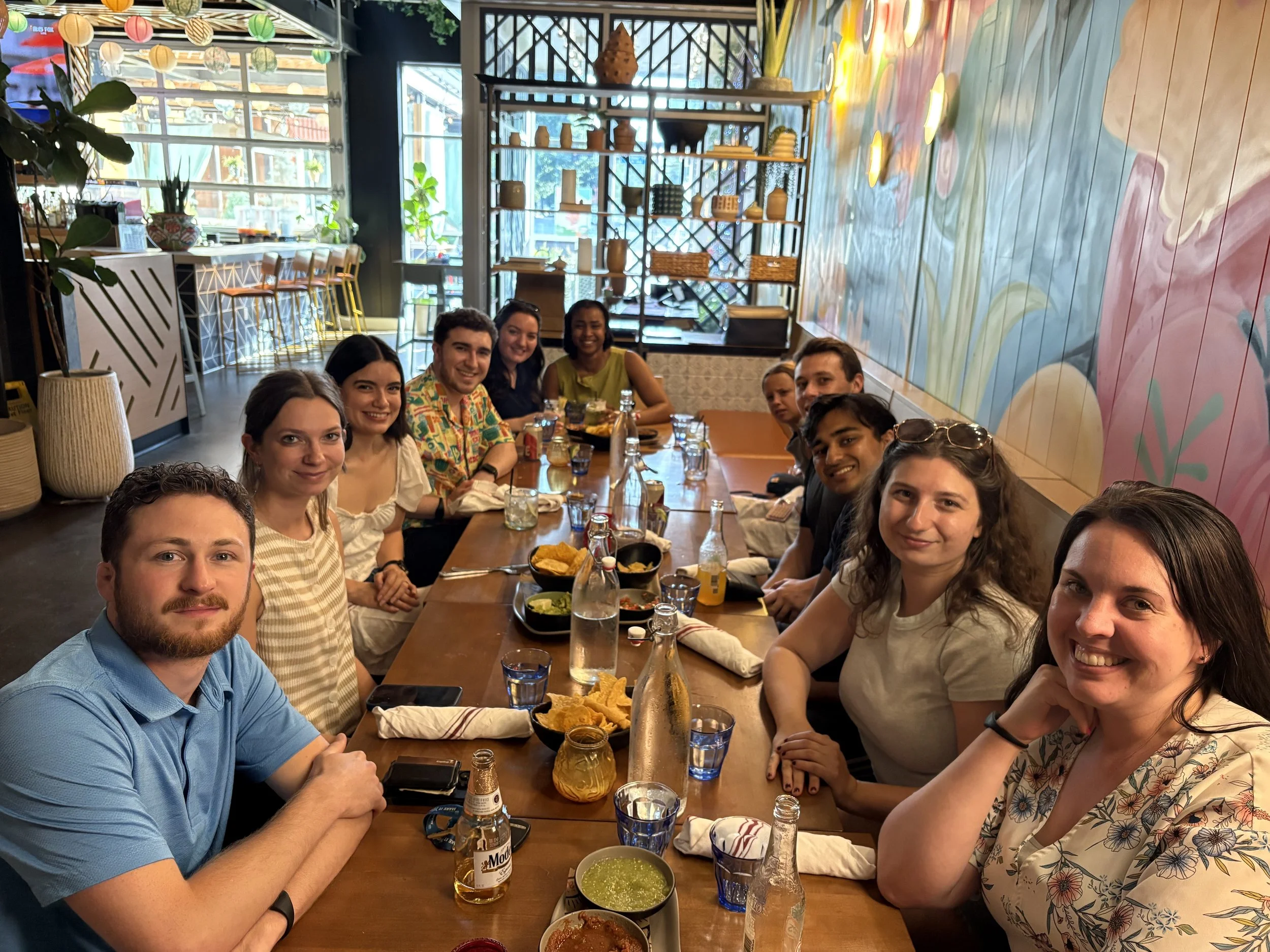
Some of the lab out celebrating the newly crowned Master Wheeler!


Written by Gia Romano

Welcome to the Hartman Lab, Tilly May!

Earlier this year our lab welcomed its newest member, Tilly May!

Tilly is an undergraduate student in the Dietrich School of Arts & Sciences and will be working with us as lab support within our BSL-2 facilities. She is currently learning cell culture, reagent aliquoting, and other basic techniques along with assisting in day-to-day lab maintenance with Kim and Jackson. Here is what Tilly had to say about what brought her to Pitt and the Hartman lab, and her passions both in and out of the lab:

“Hi! My name is Tilly May and I am from Lower Merion, PA, about 15 minutes outside of Philadelphia. I chose to attend the University of Pittsburgh because of its central urban location and specialty programs. I enjoyed the opportunities that growing up near a city gave me, and I was particularly drawn to how Pitt too offered impressive access to research experience. As a Pre-Veterinary student, I found that the city had many animal hospitals for me to get involved with clinical experience, as well as a strong science program offering rigorous coursework for pre-requisites. Going into college, I knew that I wanted to get involved with research early so that I would have the best opportunity to grow experience and responsibility throughout my four undergraduate years. To best suit my goals of vet school, I hoped to find a lab within the School of Public Health that focused on transmitted disease, and once I got in contact with the Hartman Lab I knew it was the best fit for me!”

“During my undergraduate years, I plan to continue involvement in research, as well as taking on clinical hours at a local emergency animal hospital. Throughout my summer and winter breaks I will continue shadowing and volunteering at veterinary hospitals and clinics, and have been involved in course work research and testing with the University of Pennsylvania’s Veterinary Anatomy department. After my undergraduate years, I hope to attend vet school and pursue my interest in small animal veterinary medicine.”

“When I’m not in the lab, in class, or studying, I spend a lot of my time running! Sports were a huge part of my development, giving me a decompressing outlet, and something that I knew was important for me to continue throughout college! I’m running my first half-marathon in May and planning to run the Philadelphia Marathon in November!”

Tilly learning to aliquot reagents using a Multichannel Pipette!

Welcome to the Hartman lab family, Tilly, we’re excited to have you with us!


Written by Gia Romano

Dr. Connors' OROV Neuro Paper is Published!

Another one of Dr. Kaleigh Connors’ studies has just been published, that’s two published articles in two months! Appearing in the February edition of PLOS Pathogens is the culmination of Dr. Connors’ work on characterizing the replication of historic and contemporary isolates of Oropouche virus (OROV) in neural cells. Entitled “Neural cells are susceptible to historic and recently emerged Oropouche virus strains,” the paper features work from multiple former members of our group across many model systems to elucidate the neuropathogenic potential of OROV.

OROV is a bunyavirus that is distantly related to RVFV. OROV is notable because it has caused a substantial regional outbreak from 2023-2025 in South and Central America. There were significant numbers of cases of people who developed serious neurological issues (Guillain-Barre syndrome) and brain developmental issues during pregnancy (microcephaly).

This study addresses the gap in published knowledge on OROV’s ability to infect cells of the central nervous system (CNS). Using immunofluorescent microscopy to detect viral antigens, we demonstrated that the historical strain of OROV (called BeAn19991 and was isolated in 1960) infects and replicates within immortalized neurons, microglia, and astrocytes.

Immunostained primary rat cortical neurons

Immunostained primary rat cortical neurons at 24HPI.

Permissivity of primary neural progenitors to OROV-BeAn19991 infection was also demonstrated in both human induced pluripotent stem cell-derived neuroprogenitor cells and primary rat cortical neuron cultures. These are physiologically relevant models more accurately representing the state of OROV infection of neural cells in vivo. We also utilized two emergent viral strains (OROV-AM0088 and OROV-240023), both of which were obtained during the 2024 outbreak. Although cells were slightly less susceptible to OROV infection by these strains, the results demonstrate the potential for OROV infection across host species and viral strains.

OROV replicates to high titers in human induced pluripotent stem cell-derived neuroprogenitor cells!

And in hiPSC-derived neurons too! Ooooo how pretty!

Building off Dr. Connors’ previous work with Rift Valley fever virus, we established rat ex vivo brain slice cultures as a model for neurovirulent infection of bunyaviruses. This same model was used to demonstrate viral growth kinetics and immune response in a more approximately intact CNS infrastructure. The results from all three OROV strains indicate local inflammatory responses (induction of type I interferons IFN-α and IFN-β, cytokine IL-1β, and the chemokine MCP-1).

Look at all of that inflammatory response!

Collectively, these findings provide strong evidence that both historical and newly emerged OROV strains are neurotropic and capable of replicating in diverse neural cell types. By establishing in vitro and ex vivo CNS models for OROV infection, we lay essential groundwork for future neuropathogenesis and immune response work in OROV and similar bunyaviruses. Exceptional work, Dr. Connors!


Written by Gia Romano

News blogger update!!

Gia Romano is going to be updating our lab news and website going forward! She’s been a public health undergraduate student with us. Please welcome her entries! THANKS GIA!!!

Rachael and Gia co-cell-culturing

Also check out our publications list because we have Connors x 2 in 2026 already….

Journal of Virology - Cell Death in Neurons


For the past while now, Kaleigh has been ghost writing our website News entries. I only say “ghost writing” because we couldn’t figure out how to add her name to the entries. Well you are now (temporarily) back to me (Amy) because Kaleigh has taken a job as a medical writer with Fingerpaint Group! Science writing has always been an interest of hers, and she even did an internship with UPMC media relations. Kaleigh has a couple of publications that we are wrapping up, and one was just published this week. This manuscript is the completion of a major part of her thesis work. This was a TON of fantastic work! Here is her write up of the study:


*undisclosed location


I’m so excited to share that one of the final chapters of my dissertation work is out now in the Journal of Virology! We demonstrate that Rift Valley fever virus (RVFV) results in the activation of multiple cell death pathways in neurons – a critical and fragile target of viral infection in the brain!

Using both wildtype and attenuated strains of RVFV, we used immunoblotting and fluorescent microscopy to describe which pathways of cell death were activated in primary neurons during infection. The answer: several!

    • Using whole brains obtained at endpoint in a RVFV encephalitis model, we show the activation of proteins associated with apoptosis, necroptosis, and pyroptosis. This supports findings by our group and others demonstrating the susceptibility of neural cells to RVFV infection.
    • Next, moving into a primary model using rat cortical neurons, we demonstrate that RVFV infection by both wildtype (strain ZH501) and an attenuated strain (RVFV-delNSsNSm) results in robust viral replication and subsequent neuronal damage and death.
    • We confirm the formation of activated caspase 3 filaments in the nuclei of RVFV-infected neurons that can be detected using immunofluorescent microscopy, even at 6 hpi. This aligns with the detection of RVFV-NSs filaments at the same timepoints.
    • At 6, 12 and 24 hpi we demonstrate activation of proteins associated with apoptosis, necroptosis, and pyroptosis following infection with wildtype and attenuated RVFV, including cleaved caspase 3, gasdermin E, and phosphorylated-MLKL.
    • To add to these results, we used a technique called In Cell Western (ICW) to perform more high-throughput analysis of protein activation. Compared to mock-infected neurons, we observed an increase in proteins associated with apoptosis, necroptosis, and pyroptosis.
    • Our final goal was to see if we could promote neuron survival, and reduce RVFV infection, using cell death inhibitors. We tested pan-caspase inhibitor Z-VAD-FMK and caspase 3 inhibitor Z-DEVD-FMK against wildtype RVFV and found that both reduced the activation of caspase 3 in a dose-dependent manner. However, there was not impact on viral replication at 24 or 48 hpi.

Collectively, these findings show that in primary rat neurons, infection with RVFV activates multiple mechanisms of cell death - even in the presence of viral interference - highlighting the redundancy and cross-talk of these mechanisms. This study adds to our understanding of RVFV neurologic disease, and lays the groundwork for the development of targeted therapeutics that may be both neuroprotective and antiviral.

I am immensely proud of this work (which began in 2021!), and grateful to all of my co-authors and colleagues who have seen this project through. Hartman lab members Zach Frey and Matt Demers, my co-neurovirologists; Reed lab members Morgan Midgett, Connor Wiliams and Doug Reed supported the animal work; Zak Wills from the Department of Neurobiology who provided resources, techniques, and invaluable insight into this project; and of course my mentor Amy Hartman, without whom I would’ve never dove into RVFV infection in neurons!

Read the full article here: https://doi.org/10.1128/jvi.01742-25

Kaleigh

Figure 1 - RVFV & PAN-optosis pathways

New publication in JVI!

We have a new publication from our group, led by our former undergraduate student/technician Zach Frey, now published in the Journal of Virology! This is the culmination of work across multiple folks from our group, Dr. Zak Wills in the Department of Neurobiology, and our collaborators in the Leung and Amarasinghe labs at Wash U St. Louis entitled “LRP1 facilitates Jamestown Canyon virus infection of neurons.”

While this project started as a basic characterization of Jamestown Canyon virus (JCV) neuropathogenesis, Zach found that the low-density lipoprotein receptor related protein 1 (Lrp1) plays a significant role in viral entry (see RVFV and OROV).

JCV is an orthobunyavirus endemic to North America that manifests as asymptomatic or a mild febrile illness in people. However, like other bunyaviruses, JCV infection can progress to neuroinvasive disease, an outcome that remains understudied. Incidentally, LRP1 is highly expressed in the brain, including neurons, astrocytes and microglia. When these cells (microglia, BV2; neurons, N2a) lack Lrp1, JCV binding, internalization and replication are all reduced:

The dependence on Lrp1 for infection in was further confirmed by incubating JCV with Lrp1 proteins. Zach found that after using this JCV:Lrp1 mixture to infect cells, the amount of virus collected after 24 hours was significantly reduced in a dose-dependent manner. In addition, our collaborator Dr. David Price (Leung Lab), characterized the binding affinity to these clusters:

Zach then began to characterize JCV infection in a primary neuron model of infection. At different infection doses, he found that JCV replicates well in these cells and to high titer. In addition, he confirmed that these primary neurons have Lrp1 expression. The next step was to determine if Lrp1 played a role in JCV infection of primary neurons.

Using the high-affinity LRP1 binding protein mRAP-D3, he found that he could reduce JCV infection in primary neurons in a dose dependent manner.  There is a striking reduction in JCV nucleoprotein by immunofluorescent staining at the highest dose of mRAP-D3:

Another interesting part of this story involves RVFV glycoprotein Gn. We previously identified that RVFV Gn binds to clusters II and IV of LRP1, and that treatment of cells with purified Gn protein completely inhibits infection with RVFV and OROV. Performing this experiment using JCV, one measure that Zach used was western blot of viral proteins. Zach determined that increasing concentrations of RVFV Gn competitively inhibits the accumulation of JCV nucleoprotein in the cells. Alongside his other data, this suggest that JCV likely binds to similar epitopes of Lrp1 clusters II and IV to RVFV.

Finally, since JCV infection was not completely blocked by depletion or inhibition of Lrp1 use, Zach investigated the role of other LDLR receptors. He did this by treating LRP1 knockout cells with mRAP-D3, which not only binds to LRP1, but also other LDLRs. The premise was that if JCV requires both Lrp1 and another LDLR, the combination of Lrp1 KO cells and treatment with mRAP-D3 would further reduce JCV infection. Indeed, this is what Zach found, suggesting that another LDLR may be involved in JCV entry.

These findings offer a significant advance in the understanding of viral entry and bunyavirus pathogenesis, as previous findings by our groups and others on RVFV, OROV, and SFTSV demonstrate LRP1 as a pan-bunyaviral host factor. This offers an opportunity for the development of broad pan-bunyaviral therapeutics.

Zach is currently a PhD student at UNC. This work was supported by postdocs Kaleigh and Rachael, and PMI PhD candidate Cade!